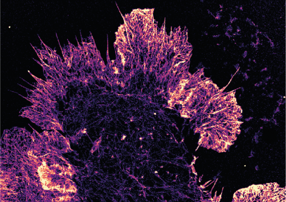
Supercharge your microscope: researchers share guide for ultra-precise 3D imaging
UNSW Sydney researchers have shared step-by-step instructions to empower other scientists to enhance the resolution and stability of single-molecule microscopes.
UNSW Sydney researchers have shared step-by-step instructions to empower other scientists to enhance the resolution and stability of single-molecule microscopes.

Isabelle Dubach
Media and Content Manager
+61 432 307 244
i.dubach@unsw.edu.au
Researchers will be able to build ultra-precise microscopes to visualise and explore the interactions between individual molecules within cells, thanks to a system made available to the scientific community by UNSW medical researchers.
Their system simply and practically overcomes the challenges associated with movement during imaging, exceeding current limits of super-resolution microscopes.
When the sample or the microscope set-up moves during imaging, errors are introduced that degrade molecular resolution – this is called drift.
“Drift is a major barrier to achieving resolution beyond the 20–30 nm set by Nobel-prize winning super-resolution fluorescence microscopy,” says Scientia Professor Katharina Gaus from UNSW Medicine’s Single Molecule Science.
“The longer it takes to image a sample, the more drift there will be. The biggest cause in drift is vibrations from people walking by, or cars driving outside the building,” she says.
Prof. Gaus explains that for single-molecule imaging, researchers typically label molecules with fluorescent dyes and get them to blink on and off using lasers.
“We can’t image them all at the same time. So, when the sample drifts on the microscope then the position of the glowing molecules at the beginning of the experiment will be different from the position at the end of the experiment, introducing an artefact,” she says.
The active stabilisation system the team of biophysicists at UNSW developed tackles this issue by adding sensors to the microscope with a feedback system to re-align the optical path when it detects the slightest change. The stabilization system automatically returns the optical path to within one nanometre of its original position in all three dimensions continuously while the samples are being imaged.
After outlining the design of their autonomous feedback system in a Science Advances publication earlier this year, the team now describe in Nature Protocols how to implement a stabilisation system to actively correct the alignment of super-resolution microscopes and eliminate drift.
“It is a how-to guide for using our feedback system on different setups. We implemented it on a range of systems, including on commercially available microscopes,” says Dr Simao Pereira Coelho, who led this project.
The protocol is designed to enable even users without specialist optical training to upgrade existing microscopes, including a guide for using the software and integrating the hardware onto a custom built or standard microscope.
“We can now image for as long as we want, to get more information out of one sample – without compromising the quality of the data. Not only does this make experiments more precise, but it opens up this new idea that you can run this completely autonomously,” says Prof. Gaus.
“The same approach can also be used in other instruments that require high precision, for example in atomic force microscopy or DNA sequencers, or where servicing and realigning an instrument manually is not so straightforward,” she says.